
Leg Bones Diagram
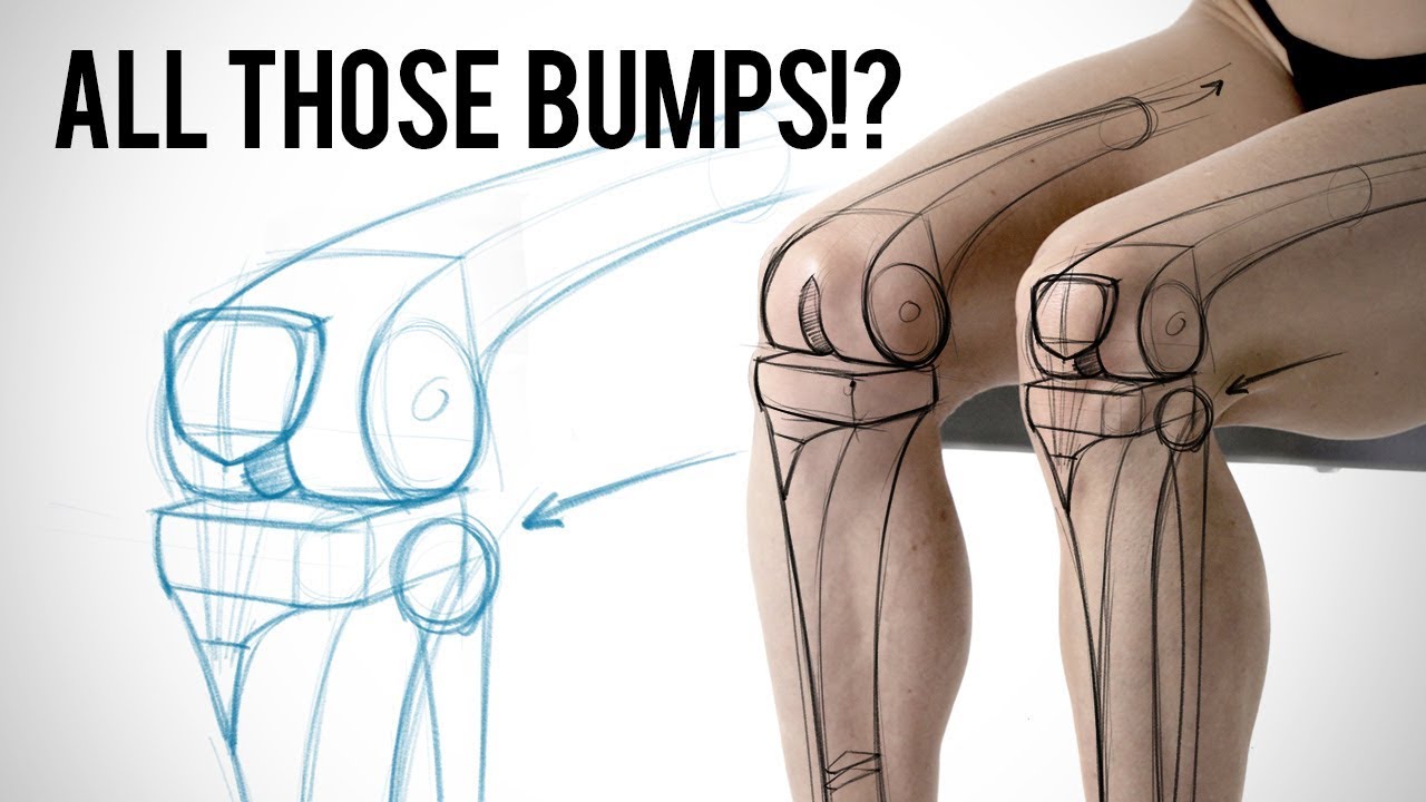
Leg Bones Diagram. When you stand or walk, all the weight of your upper body rests on them. Quizzes on human skeletal system anatomy, bone anatomy, and bone markings. He leg's main function in the human is for locomotion and support of the rest of the body. Each leg is made up of four bones. Most of the leg skeleton has bony prominences and margins that can be palpated and some serve as anatomical landmarks that define the extent of the leg.
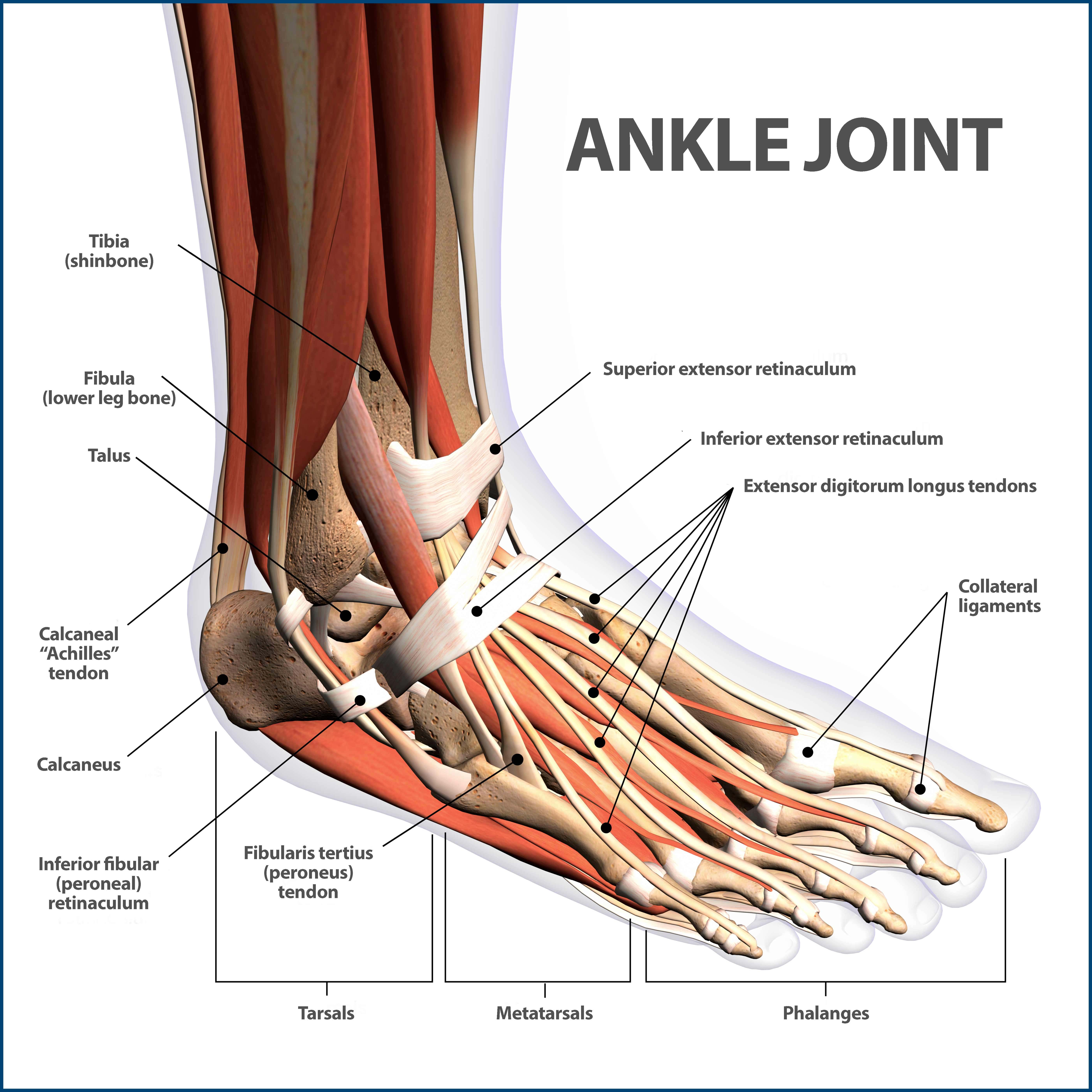
The foot bones shown in this diagram are the talus, navicular, cuneiform, cuboid. Pngtree offers bone diagram png and vector images, as well as transparant background bone diagram clipart images and psd files. Includes leg (femur, tibia, patella, and fibula) and foot (tarsals and digits) bones. Click now to learn more about the bones, muscles, and soft tissues tibia: The bones of the leg are the femur, tibia, fibula and patella.
Blood vessels and nerves enter the bone.
Health diagram bone skeleton leg knee science anchor chart human human body. The foot bones shown in this diagram are the talus, navicular, cuneiform, cuboid, metatarsals and calcaneus. Visit kenhub for more skeletal system quizzes. Click now to learn more about the bones, muscles, and soft tissues tibia: High quality realistic skeleton legs.
The human leg consists of 8 bones, 4 per leg. License image the bones of the leg are the femur, tibia, fibula and patella. Diagram of blood and nerve supply to bone. Health diagram bone skeleton leg knee science anchor chart human human body. Its lower end helps create the knee joint.
Pngtree offers bone diagram png and vector images, as well as transparant background bone diagram clipart images and psd files.
Quizzes on human skeletal system anatomy, bone anatomy, and bone markings. Diagram of blood and nerve supply to bone. Diagram and names of leg bones, diagram of foot and leg bones, diagram of leg bones, diagram of lower leg bones, diagram of the related posts of diagram of leg bones. At the microscopic level, this hard outer. The bones involved in it, however, are only the femur and the tibia, although the smaller bone of the leg, the fibula, is carried along in the movements of flexion, extension, and slight rotation that this joint.
When you stand or walk, all the weight of your upper body rests on them. Download the free graphic resources in the form of png, eps. Time to jump right into the biggest and strongest bones in the human body. Learn vocabulary, terms and more with flashcards, games and other study tools. The musculoskeletal segment of the leg, including the foot bones (ankle, heel bone, toe bones), fibula and tibia, knee, femur and femoral neck, hip and sacrum as well as the third, fourth.
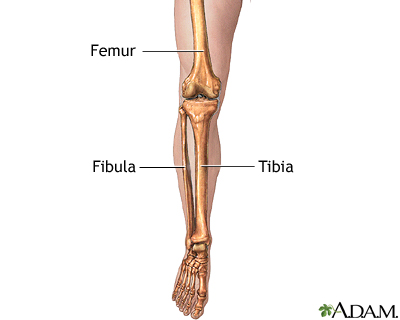
These simple labelled diagrams of the bones of the lower legs and feet and the bones of the arms and hands this diagram shows the skeletal structure of the leg (anterior view) and foot (dorsal view).
Diagram of blood and nerve supply to bone. He leg's main function in the human is for locomotion and support of the rest of the body. At the microscopic level, this hard outer. Blood vessels and nerves enter the bone. High quality realistic skeleton legs.
Posting Komentar untuk "Leg Bones Diagram"